Cellular Imaging Facility – CIF
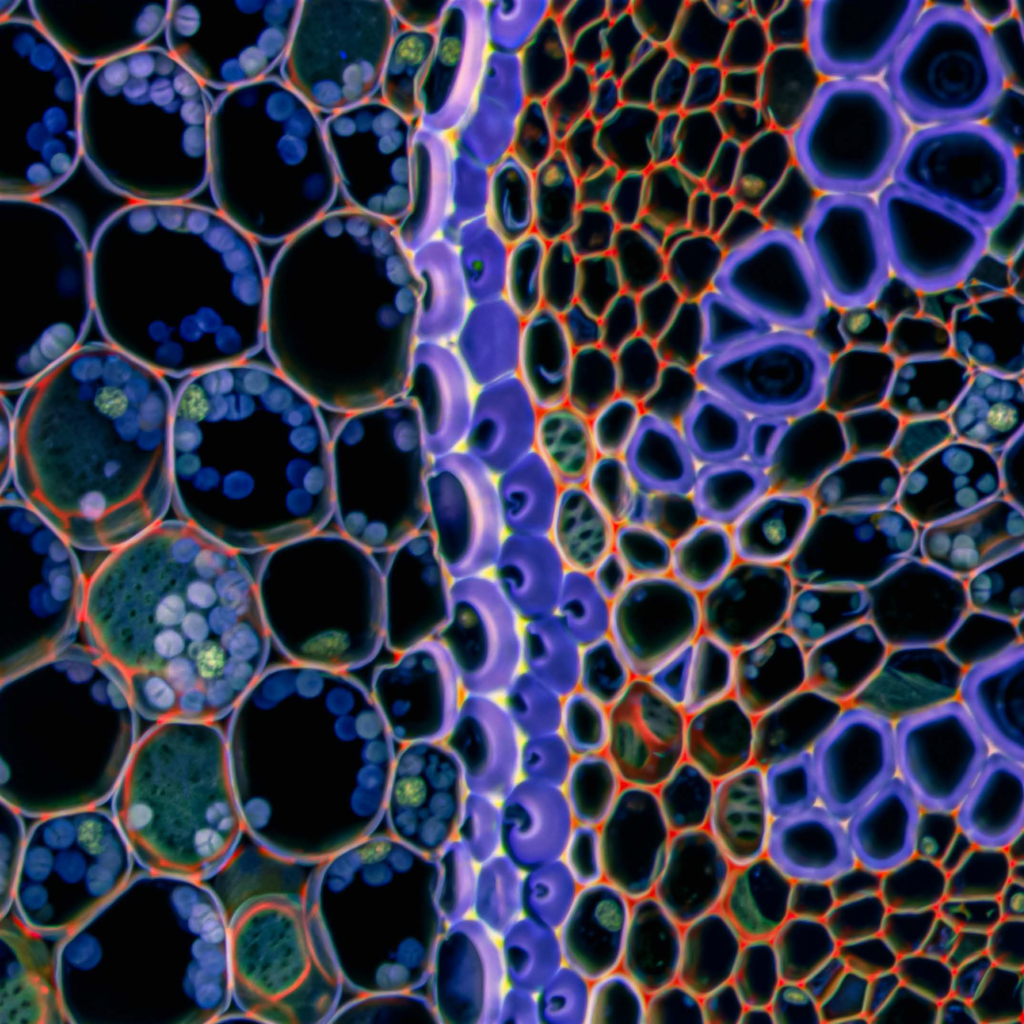
The CIF platform, created in 2003 thanks to a partnership between UNIL and the CHUV, is currently composed of four antennas located at Bugnon 9 (CIF Bugnon), at Dorigny (CIF Dorigny), on the Epalinges site (CIF Epalinges) and, as of 2018, in the AGORA research centre (CIF AGORA). The mission of the CIF platform is to meet the needs of researchers in the field of optical imaging, covering wide-field optical microscopy (fluorescence and transmission), confocal microscopy, ion imaging and videomicroscopy, as well as the processing and analysis of digital images.

Mission
The CIF has several missions and services:
- Access to a range of state-of-the-art instruments and techniques is offered to researchers from AGORA and associated institutions.
- The CIF shares and disseminates the theoretical and practical know-how of these approaches through different teaching modes (training in the handling of instruments throughout the year, an introductory course in cell imaging, a series of specialised modules on various aspects of cell imaging in the form of workshops).
- In parallel to the services provided, the CIF, together with a consortium of interested researchers, develops and implements some advanced optical and imaging technologies.
Resources
The CIF provides users with a wide range of image acquisition and processing instruments, which are accompanied by individualised practical training.
List of available resources at the CIF Agora branch:
- Olympus FluoView 3000 – point scanning confocal
Applications: Confocal acquisition, 3D fluorescence reconstruction, optimal resolution, acquisition of large areas, colocalization.
- Nikon Ti2 | Yokogawa CSU-W1 – spinning disk confocal
Applications: live-cell imaging, single multi-well plate imaging, confocal acquisition, fast acquisition of 3D volumes and larges areas.
- Zeiss Axio Observer Z1 – Bright-field and widefield fluorescence microscope
Applications: General immunofluorescence, histology / stained slides, large area acquisition
- 3 Image analysis and processing workstations
Software available: IMARIS (Cancer Research package + Coloc + Filament Tracer + Cell + XT), Imagej/FIJI, QuPath, CellProfiler, NIS-elements AR Analysis, NDP.view2.
Access to Huygens Remote Manager for deconvolution.
- Additional resources are available on the close-by CIF branches:
e.g. slide scanners, light-sheet microscope for clarified tissue, laser-capture microdissection systems


